EARLY DIAGNOSIS OF ORAL CANCER WITH FLUORESCENCE: CLINICAL CASE
Prof. Silvio Abati
Associate Professor of Odontostomatological Diseases University Vita-Salute San Raffaele of Milan
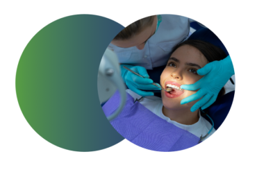
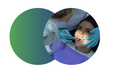
Early detection of cancer oral squamouscell, the cancer that originates from the epithelium of the oral mucosa, reduces morbidity and increases the survival of patients. Histopathological analysis of suspected lesions is the gold standard for the precise diagnosis of oral cancer. The finding of lesions of the premalignant mucosa or malignant, initial and small requires more clinical experience and is sometimes more complex. This is fundamental for the anticipation of the diagnosis of cancer, both in patients who had never manifested suspected lesions of the mucosa and in patients already treated for oral cancer and in follow-up. Scientific evidence (1-4) shows that screening for oral cancer with conventional visual examination can reduce mortality in highrisk individuals. Optical aids with good sensitivity and acceptable specificity can be used in combination with the conventional visual examination. The detection of tissue autofluorescence of the oral mucosa is carried out by dentists with portable devices that allow the illumination of the mucosa with short-wave or UV blue light, between 380 and 480 nm. Healthy oral tissues emit a characteristic green fluorescence after illumination of excitation and premalignant injury zones. Dysplasia or malignant (carcinoma) are highlighted as dark areas for the loss of autofluorescence linked to tissue phenomena and metabolic in the areas of injury.